Steps Of Placental Examination
In the next step placental examination is suggested in all cases together with molecular cytogenetic evaluation and. The clinical history is crucial in placental assessment.

Duodenum Sonography Student Descriptive Education
Up to 12 cash back Placental examination.
Steps of placental examination. Stepwise Standardized Approach to the Basic Obstetric Ultrasound Examination 101in the Second and Third Trimester of Pregnancy. The sequence is fetal presentation heartbeat number of fetuses placental position amniotic fluid estimation biometry 3. Visit 1 Day 1.
If positive it indicates TB infection and a chest x-ray and further evaluation is necessary. Steps to examining the placenta. The addition of autopsy resulted in 78 74 cases having an identifiable probable cause of death.
An important first step in the analysis of PD is to identify the case circumstances including relevant details regarding maternal history obstetric history and current pregnancy complications are evaluated and recorded. Records were reviewed in a stepwise manner. Palpate the lower uterus below the umbilicus to find the presenting part.
Thirty minutes prior to the onset of back pain she noted. Standardisation enhances trainees confidence. 6 Completing the Examination.
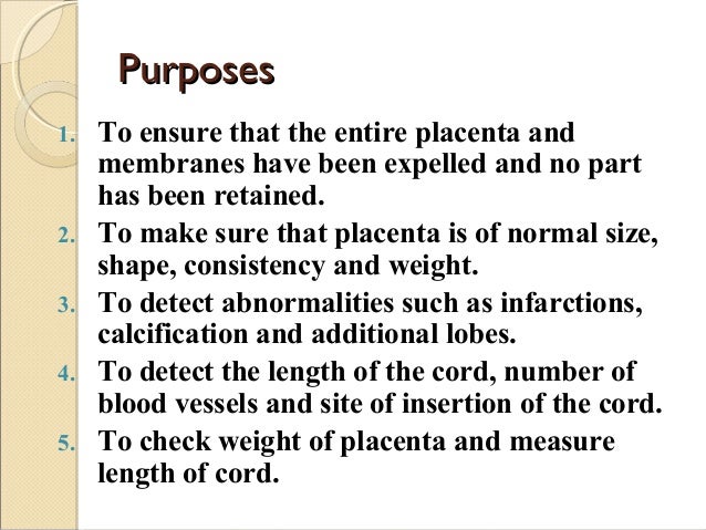
To achieve optimal benefit from placental reports it is essential to standardise the method of placenta examination. The doctor or midwife will look for missing pieces the shape and consistency of your placenta. The sequence is fetal presentation heartbeat number of fetuses placental position amniotic fluid estimation biometry 3.
Classification of placental abruption is based on extent of separation ie partial vs complete and the location of separation ie marginal vs central. This article summarises the clinical indications for placenta referral and the most common acknowledged clinicopathological correlations. Visit 2 Day 3.
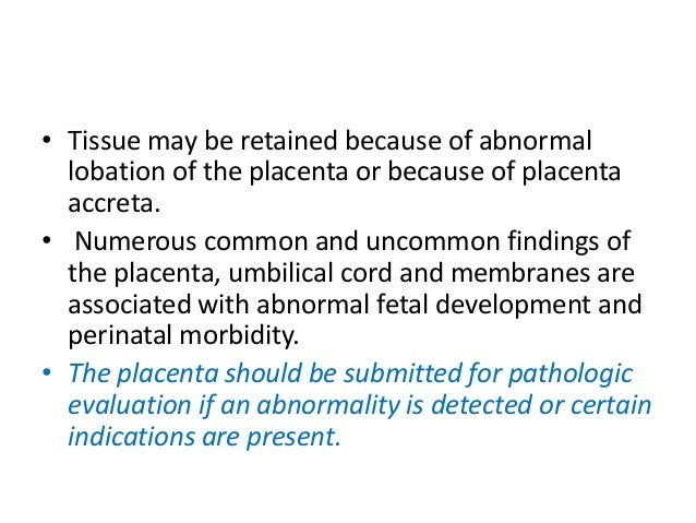
Thus while the placental weight or villous histology may be normal for. External examination Photographs Surface Swabs XRAYUltrasound Post-mortem examination YN Cord blood not possible for most stillbirths Full Blood Count Cord gases Placenta Cord see page 5 for details Macroscopic examination of placenta and cord Microbiological Cultures swab between chorion and amnion Cord and placental section for. After placental pathologic examination 88 61 cases had a probable cause of death identified.
This video demonstrates the methods for examination of the placenta. PPD test is read within 48-72 hrs. At each step a cause of death and the certainty of that etiology were coded.
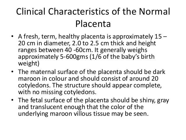
Usually in the center of the fetal side side that faces the baby in the womb. They will look at how the cord inserts into the placenta and whether or not there are calcifications. Identify placental or fetal conditions that can be recurrent or inherited.
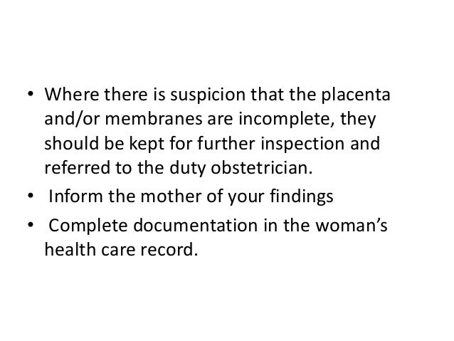
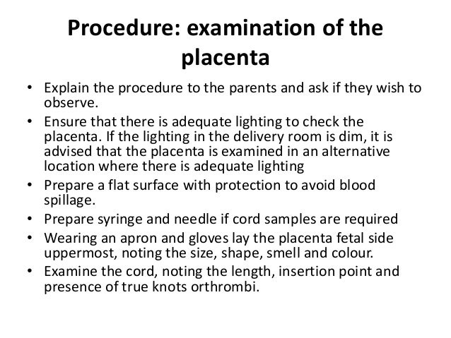
Step 1 Accoucheur examination of the placenta membranes and cord using sterile gloves Cord insertion Circle Eccentric Central Marginal Velamentous Other. Examination of the placenta Explain the procedure to the parents and ask if they wish to observe. For example the gestational age of the fetusneonate is important as the placental disc weight increases over gestation and placental villi mature over time.
Autopsy led to additional clinical management changes in 6 6 cases. Note the cord insertion. If breech presentation is suspected the fetal head can be often be palpated in the upper uterus.
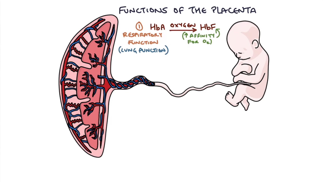
- Fetal lie and presentation - Fetal cardiac activity - Number of fetuses in the uterus - Adequacy of amniotic fluid - Localization of the placenta - Fetal biometry. There are also tests that may be run on the placenta including ones to look for diseases or infections. Overview of Placental Exam.
First the clinical history and laboratory results then the placental pathologic evaluation and finally the autopsy. Firm and round signifies cephalic soft andor non-round suggests breech. A 30-year-old G1P0 woman at 36 weeks of gestation presents to the emergency room with sudden onset of moderate back pain and strong uterine cramping that began 2 hours ago.
Standardisation enhances confidence in trainees. 3 Abdominal Examination. The following is the standard process for completing a two-step TB PPD test.
Automated placenta analysis based on photographic imaging may allow more placentas to be examined reduce the number of normal placentas sent for full pathological examination allow for timely on-site initial diagnosis and provide an objective morphological and pathological analysis. Ultrasound examination in the 2nd 3rdtrimester of pregnancy should be approached systematically 2. Hold the umbilical cord in one hand and let the placenta and membranes hand down like an inverted umbrella The umbilical vessels will be seen passing from the cord and gradually fading into the edge of the placenta Look for free-ending vessels and holes which may indicate that a succenturiate lobe.
Ensure that there is adequate lighting to check the placenta. Placental examination alone changed clinical management in 52 36 cases. Placental abruption is mainly a clinical diagnosis based on findings of vaginal bleeding abdominal pain uterine tenderness uterine contractions and fetal distress.
Ultrasound examination in the 2 nd and 3rd trimester of pregnancy should be approached systematically 2. If the lighting in the delivery room is dim it is advised that the placenta is examined in an alternative location where there is adequate lighting Prepare a flat surface with protection to avoid blood spillage. Two-Step TB PPD test completed within the past year.
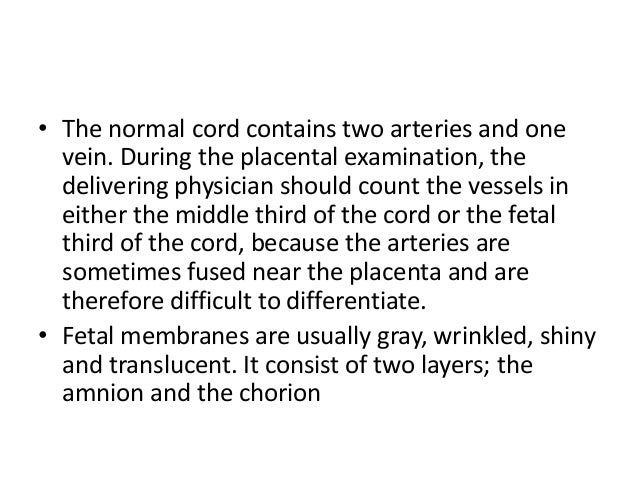
Sometimes on the side and rarely out in the amniotic membrane and joining the placenta only by blood vessels velamentous insertion. PPD antigen is applied under the skin. To automate the assessment of placentas Penn State.
Understanding The Placenta Youtube
Placental Examination Pdf Placenta Anatomy


Examination Of Placenta In Hindi Surface Of Placenta Placental Examination Nursing Anm Gnm Youtube

Plgf Placental Growth Factor Testing In Clinical Practice Evidence From A Canadian Tertiary Maternity Referral Center Hypertension

Pin By Kymberley Leveque On Labor In 2021 Midwifery Midwifery Student Pediatric Nursing
Placenta Examination

Significance Of Pathological Examination Of The Placenta With A Focus On Intrauterine Infection And Fetal Growth Restriction Nakayama 2017 Journal Of Obstetrics And Gynaecology Research Wiley Online Library
Placenta Examination

Variant Placental Morphology Download Table

Placenta Praevia Causes Clinical Features Management Teachmeobgyn

Pin On Med Students

Placental Insufficiency What Is It Causes Symptoms Treatment And More Osmosis

Placenta Ultrasound Ultrasound Sonography Ultrasound Tech Ultrasound Technician
Placenta Examination
Placenta Examination

Variant Placental Morphology Download Table
Placenta Examination

Placenta Examination
Placenta Examination


Posting Komentar untuk "Steps Of Placental Examination"